World’s first: Lung cancer surgery with Orsi Innotech’s augmented reality technology
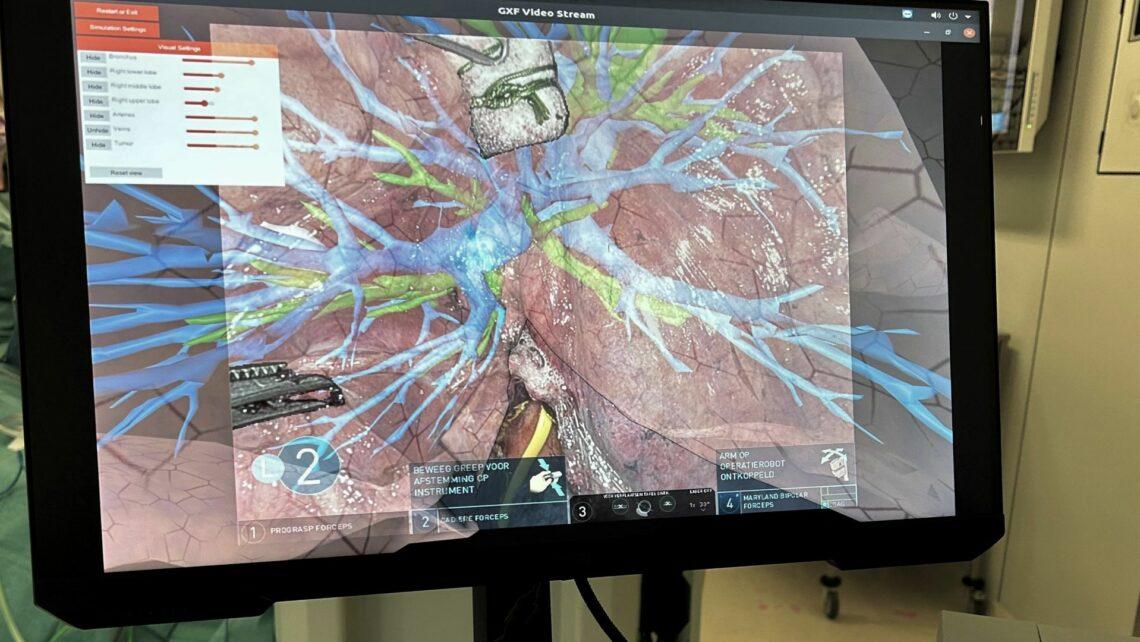
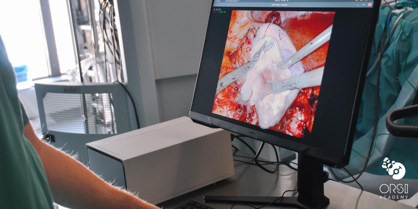
Surgeons at Erasmus Medical Center have become the first in the world to remove a lung tumor with the help of augmented reality, developed by Orsi Innotech. This technology enables them to perform the world’s first robot-assisted lobectomy using augmented reality. During this surgery, virtual overlay of the tumor, blood vessels and airways were projected over the camera image of the patient’s lung and was rendered with real-time AI-assisted robotic instrument detection. This allows surgeons to find their way inside the patient’s body more safely & effectively.

Dr. Amir Sadeghi and Dr. Sabrina Siregar of Erasmus MC (Rotterdam, The Netherlands), in collaboration with Orsi Innotech, are among the first in the world to use augmented reality in robot-assisted lung surgery. During surgery, live virtual images of the lung are projected over the camera image of the patient’s actual lung, allowing the surgeon to see crucial information, such as the location of the blood vessels and airways, as well as the exact position of the tumor.
“Compare augmented reality with a GPS,” explains researcher and heart lung surgeon-in-training Dr. Amir Sadeghi. “Augmented reality helps the surgeon operate more precisely. For example, we can now see that the exact position of the artery”. Details that cannot be seen with the naked eye can be seen with the help of augmented reality.
Augmented reality as GPS for safer surgery
Benefits
The use of augmented reality in robot-assisted surgery will lead to faster procedures and fewer patient complications. In addition, the use of this technology will enable an accelerated learning curve for novice surgeons.
Combination of patient-specific virtual and deformable lung models, developed by Dr. Sadeghi (Erasmus MC) in collaboration with MedicalVR, and augmented reality technology developed by Orsi Innotech in collab with NVIDIA & Deltacast, resulted in the first successful use cases in robot-assisted lung surgery.


